GENERAL PROCEDURES:
1. Make sure all backpacks are out of the
aisles before you get a microscope! Always carry the microscope
with one hand on the Arm and one hand on the Base. Carry it close
to your body.
2. Remove the cover, plug the microscope in, and
place the excess cord on the table! If you let the excess cord dangle
over the edge, your knee could get caught on it, and the next sound you hear
will be a very expensive crash. I will
bill you later!

3. Always
start and end with Low Power! The Green means GO! -- “Go
ahead and put the slide on the stage.” “Go ahead and use the
Coarse focus knob.” “Go ahead and remove the slide from the
stage.” “Go ahead and put the microscope away.”
4. Place the slide on the microscope stage,
with the specimen directly over the center of the glass circle on the
stage. Then you have a 9 out of 10 chance of finding the specimen as soon
as you look through the eyepiece!
5. If, and
ONLY if, you are on LOW
POWER, lower the objective lens to the lowest
point, then focus using first the coarse knob, then the fine focus
knob. The specimen will be in focus when the LOW
POWER objective is close to the lowest point,
so start there and focus by slowly raising the lens. If
you can’t get it at all into focus using the coarse knob, then switch
to the fine focus knob.
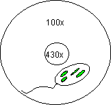
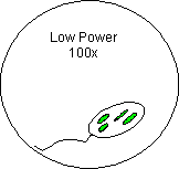
6. Once you have found the specimen on Low Power
(100x), unless specifically asked to draw it on low power, center
the specimen in your field of view, then, without changing the focus knobs,
switch it to High Power. If you don’t center the specimen you will lose
it when you switch to High Power (Yellow).
[See Above]
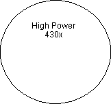
7. Once you have it on High Power remember
that you only use the fine focus knob! The
Yellow means CAUTION! -- “Caution, use
only the fine focus knob.” “Caution, do not remove the slide when it is on
High Power.” --
The High Power Objective (430x) is very close to the slide.
Use of the coarse focus knob will scratch the lens, and crack the
slide. More expensive sounds . . .
8. NEVER USE
THE RED LENS. The Red Means STOP!! -- “Stop!
Don’t use that lens!” -- It
is an oil immersion lens. Without the oil to lubricate the lens, you
will destroy it! More expensive sounds
. . . Also, the oil is needed to help gather enough light to
actually see through the lens!
Tips On Making Good Drawings:
1. Don’t even think of starting your drawing
unless you have a PENCIL! Drawings in PEN are UNACCEPTABLE! This is for
two reasons:
(a) You can erase pencil!
(b) You can shade in areas
more easily in pencil.
2. Each Drawing must be 1/2 page in size,
and must include clear, proper labels! In the upper left hand corner of
each circle include the specimen name as written on the slide label. In
the upper right hand corner, include the magnification (100x or 430x).
3. Labels should start on the outside of
the circle. The circle indicates the field of view as seen through the
eyepiece. All arrows should end with the point touching
the object to be labeled! 
4. Animal cells should always include at
least the following four labels: Cell membrane, Nuclear
membrane, Nucleus, Cytoplasm.
5. Plant Cells should always include at
least the following six labels: Cell membrane, Cell wall,
Nuclear membrane, Nucleus, Cytoplasm, Chloroplast (this last does not
exist in certain plant cells).
6. Remember: This class is about Connections!
I don’t want you to Look at the cells; I want you to SEE them!
Apply your knowledge of cell structure to your drawings! An
unlabeled drawing is nothing more than scratches on a piece of
paper!
How To Make A Wet Mount:
1. Gather a thin slice/piece of whatever
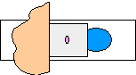
your specimen is. If your specimen is too thick, then the coverslip will
wobble on top of the sample like a see-saw:

2. Place ONE drop of water directly
over the specimen. If you put too much water over the specimen,
then the coverslip will float on top of the water, making it harder to draw
the specimens as they float past the field of view!
3. Place the coverslip at a 45 degree angle
(approximately), with one edge touching the water drop, and let go.

How To Stain a Slide:
1. Place one drop of Methylene
Blue stain on one edge of the coverslip, and the flat edge of a piece of paper
towel on the other edge of the coverslip. The paper towel will draw the
water out from under the coverslip, and the cohesion of the water (due to
those perennial favorites - Hydrogen Bonds) will draw the stain
under the coverslip.
2. As soon as the stain has covered the area
containing the specimen you are finished. The stain does not need
to be under the entire coverslip. If the stain does not cover the area needed,
get a new piece of paper towel and add more stain until it does.
3. Be sure to wipe off the excess stain with
a paper towel, so you don’t end up staining the objective lenses.
4. You are now ready to place the slide on the
microscope stage. Be sure to follow all the instructions on the previous pages
as to how to use the microscope.
5. When you have completed your drawings, be sure
to wash and dry both the slide and the coverslip and return them
to the correct places!
6. All slides must be put away in the
proper trays! Students will not leave until all materials have been
put way properly. You are a team!
Final Note:
NOTE: These procedures
will remain the same,
regardless of the type of stain,
or the addition of a hypertonic/hypotonic solution to your specimen.
REMEMBER: Be careful with
the equipment,
and be sure to leave the lab
in the same condition it was in when you arrived.